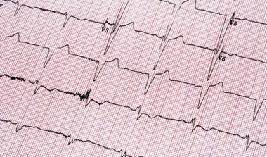
Electrophysiological Studies
What is an electrophysiological study?
An electrophysiological study (EP study) is a test used to evaluate your heart's electrical system and to check for abnormal heart rhythms.
Natural electrical impulses coordinate contractions of the different parts of the heart. This helps keep blood flowing the way it should. This movement of the heart creates the heartbeat, or heart rhythm.
During an EP study, your doctor inserts small, thin wire electrodes into a vein in the groin (or neck, in some cases). He or she will then thread the wire electrodes through the vein and into the heart. To do this, he or she uses a special type of X-ray “movie,” called fluoroscopy. Once in the heart, the electrodes measures the heart’s electrical signals. Electrical signals are also sent through the electrodes to stimulate the heart tissue to try to cause the abnormal heart rhythm. This is done so that it can be evaluated and its cause can be found. It may also be done to help evaluate how well a medicine is working.
During the EP study, specialists in heart rhythms or an electrophysiology specialist may also map the spread of the heart’s electrical impulses during each beat. This may be done to help find the source of an abnormal heartbeat.
Why might I need an electrophysiological study?
Your healthcare provider may advise an EP study for these reasons:
-
To evaluate symptoms such as dizziness, fainting, weakness, palpitation, or others to see if they might be caused by a rhythm problem. This may be done when other tests have not been clear and your doctor strongly suspects you have a heart rhythm problem
-
EP studies can be used to get information related to abnormally fast or slow heart rhythms
-
To find the source of a heart rhythm problem with the intent to do ablation once the source is identified
-
To see how well medicine(s) given to treat a rhythm problem are working
There may be other reasons for your healthcare provider to recommend an EP study.
What are the risks of an electrophysiological study?
Possible risks of an EP study include:
-
Bleeding and bruising at the site where the catheter(s) is put into a vein
-
Damage to the vessel that the catheter is put into
-
Formation of blood clots at the end of the catheter(s) that break off and travel into a blood vessel
-
Rarely, infection of the catheter site(s)
-
Rarely, perforation (a hole) of the heart
-
Rarely, damage to the heart's conduction system
For some people, having to lie still on the procedure table for the length of the study may be uncomfortable or painful.
There may be other risks depending on your specific medical condition. Be sure to discuss any concerns with your healthcare provider before the test.
How do I get ready for an electrophysiological study?
-
Your healthcare provider will explain the test to you and give you a chance to ask questions.
-
You will be asked to sign a consent form that gives your permission to do the test. Read the form carefully and ask questions if anything is not clear.
-
Tell your healthcare provider if you are sensitive to or are allergic to any medicines, iodine, latex, tape, or anesthetic agents (local and general).
-
You will need to fast (not eat or drink anything) for a certain period before the test. Your healthcare provider will tell you how long to fast, usually overnight.
-
If you are pregnant or think you may be, tell your healthcare provider.
-
Tell your provider if you have any body piercing on your chest or abdomen (belly).
-
Be sure your healthcare provider knows about all medicines (prescription and over-the-counter), vitamins, herbs, and supplements that you are taking.
-
Tell your provider if you have a history of bleeding disorders or if you are taking any anticoagulant (blood-thinning) medicines, aspirin, or other medicines that affect blood clotting. You may need to stop some of these before the test.
-
Your provider may request a blood test before the test to determine how long it takes your blood to clot. Other blood tests may be done as well.
-
A sedative (a drug to make you relax) is often given before the test, so you will need someone to drive you home afterwards.
-
Based on your medical condition, your healthcare provider may request other specific preparation.
What happens during an electrophysiological study?
You may have an EP study on an outpatient basis or as part of your stay in a hospital. Testing may vary depending on your condition and your healthcare provider’s practices.
Generally, an EP study follows this process:
-
You will be asked to remove any jewelry or other objects that may interfere with the test.
-
You will remove your clothing and put on a hospital gown.
-
You will be asked to empty your bladder before the test.
-
If there is a lot of hair at the area of the catheter insertion (often the groin area), the hair may be shaved off. This will help in healing and reduce the chance of infection after the test.
-
An intravenous (IV) line will be started in your hand or arm before the test. This is so that medicine and IV fluids can be given, if needed.
-
A member of the medical team will connect you to an electrocardiogram (ECG) monitor to record the electrical activity of your heart and monitor your heart during the test using small electrodes that stick to your skin. The team will also monitor your vital signs (heart rate, blood pressure, breathing rate, and oxygen level).
-
There may be several monitor screens showing your vital signs and the images of the catheter being moved through your body into your heart.
-
You will likely be given a sedative in your IV before the test to help you relax. However, you will be somewhat awake during the test.
-
Your doctor may check and mark your pulses below the IV site to check the circulation to the limb below during and after the test.
-
A member of the medical team will inject a local anesthetic into the skin at the site where the catheter and wires are to be put into the vein. You may feel some stinging at the site for a few seconds after the local anesthetic is injected.
-
Once the local anesthetic has taken effect, your doctor will insert a sheath, or introducer, into the blood vessel. This is a plastic tube through which the catheter(s) will be put into the blood vessel and advanced into the heart. Catheters are long, thin hollow tubes that provide a path through the blood vessels to protect the surrounding blood vessel from trauma of the equipment passing through the vessel.
-
One or more catheters will be put into the sheath and into the blood vessel. The doctor will thread the catheters through the blood vessel into the right side of the heart. Fluoroscopy (a special type of X-ray that is displayed on a TV monitor), is used to help advance the catheters to the heart. Your doctor may let you watch this process on the screen.
-
Once the catheter(s) is in the right place, your doctor will send very small electrical impulses to certain areas within the heart. You may feel your heart beat stronger and faster. If a heart rhythm abnormality is started, you may feel lightheaded or dizzy. Medicine may be given or a shock may be delivered to stop the arrhythmia. You may be sedated before a shock is given.
-
If a certain area of tissue is found to be causing a rhythm problem, the doctor may do an ablation to destroy the abnormal tissue. This is done with heat (radio waves, called radiofrequency ablation) or cooling (called cryothermy or cryoablation).
-
Sometimes adrenaline type medicines are given to help induce arrhythmia. You may feel your heart beating more rapidly and forcefully. You may feel some anxiety.
-
If you notice any discomfort or pain, such as chest pain, neck or jaw pain, back pain, arm pain, shortness of breath, or breathing difficulty, let the doctor know right away.
-
Once the EP study is done, the catheter(s) will be removed. Pressure will be put on the insertion site so that a clot will form. Once the bleeding has stopped, a very tight bandage will be placed on the site. A small sandbag or other type of weight may be placed on top of the bandage for extra pressure on the site, especially if the groin was used.
-
The staff will help you slide from the table onto a stretcher so that you can be taken to the recovery area. If the catheter was put in the groin, you won't be able to bend your leg for several hours. To help you remember to keep your leg straight, the knee of the affected leg may be covered with a sheet and the ends will be tucked under the mattress on both sides of the bed to form a type of loose restraint.
-
The results of the study may also help your healthcare providers decide whether more treatment is needed and which treatment would be best. You may need a pacemaker or implantable defibrillator, receive or change medicines, undergo an ablation procedure, or receive other treatments.
What happens after an electrophysiological study?
In the hospital
After the test, you may be taken to the recovery room for observation or returned to your hospital room. You will stay flat in bed for several hours after the test. A nurse will monitor your vital signs, the insertion site, and circulation or sensation in the affected leg or arm.
Let your nurse know right away if you feel any chest pain or tightness, or any other pain, as well as any feelings of warmth, bleeding, or pain at the insertion site.
Bed rest may vary from 2 to 6 hours depending on your specific condition.
In some cases, the sheath or introducer may be left in the insertion site. If so, you will be on bed rest until the sheath is removed. After the sheath is removed, you may be given a light meal.
After the specified period of bed rest, you may get out of bed. The nurse will help you the first time you get up, and may check your blood pressure while you are lying in bed, sitting, and standing. You should move slowly when getting up from the bed to avoid any dizziness from the long period of bed rest.
You may be given pain medicine for pain or discomfort related to the insertion site or having to lie flat and still for a prolonged period.
You may go back to your usual diet after the test, unless your healthcare provider tells you otherwise.
When you have recovered, you may be discharged to your home unless your doctor decides otherwise. If this test was done on an outpatient basis, you must have another person drive you home.
At home
Once at home, check the insertion site for bleeding, unusual pain, swelling, and abnormal color or temperature change. A small bruise is normal. If you notice a constant or large amount of blood at the site that can’t be contained with a small dressing and stopped by putting pressure over the area, contact your doctor right away.
It will be important to keep the insertion site clean and dry. Your doctor will give you specific bathing instructions.
You may be advised not to participate in any strenuous activities for a few days after the test. Your doctor will tell you when you can return to work and go back to your normal activities.
Contact your healthcare provider if you have any of the following:
-
Fever with a temperature higher than 100.4°F (38.0°C) or chills
-
Increased pain, redness, swelling, or bleeding or other drainage where the catheter was inserted
-
Coolness, numbness or tingling, or other changes in the affected leg
-
Chest pain or pressure, nausea or vomiting, profuse sweating, dizziness, or fainting
Your healthcare provider may give you other instructions after the test, depending on your situation.
Next steps
Before you agree to the test or the procedure make sure you know:
-
The name of the test or procedure
-
The reason you are having the test or procedure
-
What results to expect and what they mean
-
The risks and benefits of the test or procedure
-
What the possible side effects or complications are
-
When and where you are to have the test or procedure
-
Who will do the test or procedure and what that person’s qualifications are
-
What would happen if you did not have the test or procedure
-
Any alternative tests or procedures to think about
-
When and how will you get the results
-
Who to call after the test or procedure if you have questions or problems
-
How much will you have to pay for the test or procedure


